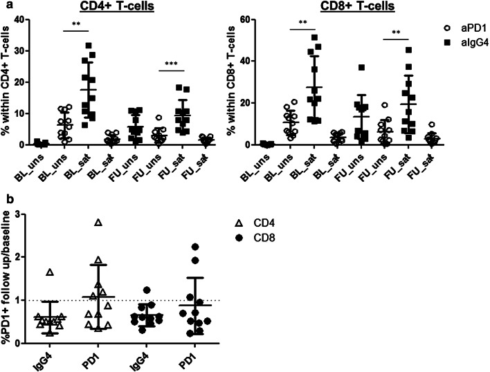
Fig. 2.
a Cryopreserved PBMCs from n = 11 melanoma patients undergoing pembro/nivo treatment were obtained before (baseline, BL) and 42 days (median) after the first anti-PD-1 injection (follow up, FU). PBMCs were stained with anti-IgG4 antibody followed by staining of surface PD-1 using clone MIH4. Prior to this staining, samples were incubated either with pembro/nivo (saturation step, sat) or buffer (unsaturated, uns). Mean percentages + SD of PD-1+ cells, detected directly (white circles) or indirectly (black squares) within CD4+ (left) and CD8+ T-cells (right) are displayed. Asterisks indicate results from t test (*p ≤ 0.05, **p ≤ 0.01, ***p ≤ 0.001). b Alterations to PD-1 expression during treatment were evaluated by calculation of fold changes at follow up/baseline. Mean fold changes + SD, detected directly (PD-1) or indirectly (IgG4) within CD4+ (white triangles) and CD8+ T-cells (black circles) are displayed

