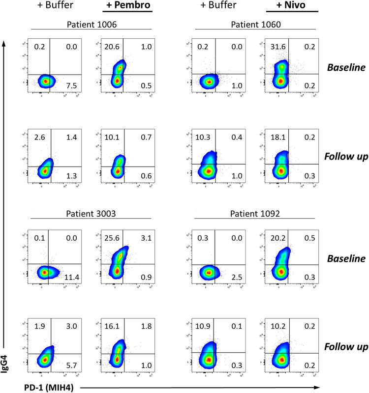
Fig. 3.
Examples of stainings from four melanoma patients undergoing pembro (left) or nivo (right) treatment were obtained before (baseline) and 42 days (median) after the first anti-PD-1 injection (follow up). PBMCs were stained with anti-IgG4 antibody followed by staining of surface PD-1 using clone MIH4. Similar results were obtained using the EH12 clone antibody (Supplementary Figure 1). Prior to this staining, samples were incubated either with buffer, pembro or nivo (saturation step). Signals from the anti-PD-1 antibody (x-axis) and the anti-IgG4 antibody (y-axis) within all viable CD8+ T-cells are displayed. All gates were placed according to corresponding Isotype control-stained cells

