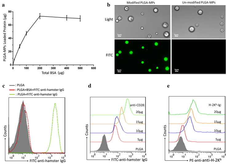
Fig. 2.
Capacity of PLGA-MPs to couple proteins. a The amount of BSA coupled onto the surface of PLGA-MPs (5 × 10⁶) was detected via micro-BCA protein assays. b FITC-anti-hamster IgG was coupled onto the surface of PLGA-MPs and detected with a fluorescence microscope. Green fluorescence was observed on the surface-modified PLGA-MPs, but not on the un-modified PLGA-MPs. c Surface-modified PLGA-MPs were incubated with FITC-anti-hamster IgG before (green) and after (red) blocking with 30% BSA and then detected by flow cytometry. Furthermore, PLGA-MPs (1 × 10⁸) were incubated with the indicated amounts of anti-CD28 or H-2Kb-Ig overnight, blocked with BSA, and then stained with FITC-anti-hamster IgG (binds to anti-CD28) or PE-anti-H-2Kb. The fluorescence shift of FITC (d) or PE (e) was detected in a range from 5 to 20 μg of anti-CD28 or H-2Kb-Ig

