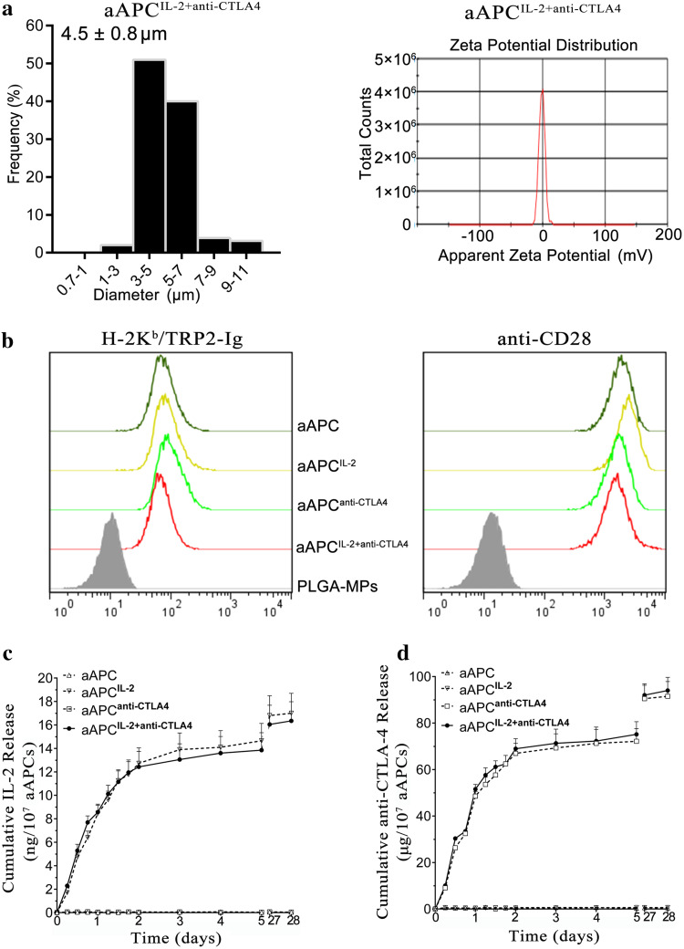
Fig. 3.
Characterization of aAPCs. a Size distribution and zeta potential distribution of aAPCIL-2+anti-CTLA4. b Phenotype analyses of aAPC, aAPCIL-2, aAPCanti-CTLA4, and aAPCIL-2+anti-CTLA4. All types of aAPCs were stained with PE-anti-H-2Kb and FITC-anti-hamster IgG to detect the co-immobilization of H-2Kb/TRP2-Ig dimers and anti-CD28 mAbs onto PLGA-MPs. Blank PLGA-MPs that were pre-blocked with 30% BSA displayed baseline staining. c, d Release profiles of IL-2 and anti-CTLA-4 from the four types of aAPCs over the course of 28 days, as detected by ELISA kits

