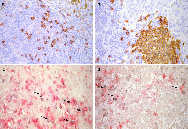
Fig. 1.
CD20+ B lymphocytes (AEC, red) dispersed in the infiltrate (a) and clustered in dense aggregate (b) in melanoma. Double staining for OX40 or CD25 (developed by Vector SG, gray signal) and CD20 (developed by fuchsin, red signal). CD20+ B cells can be seen in close contact with CD25+ (c) and OX40+ (d) T lymphocytes (arrows). Pictures were taken using ×40 objective

