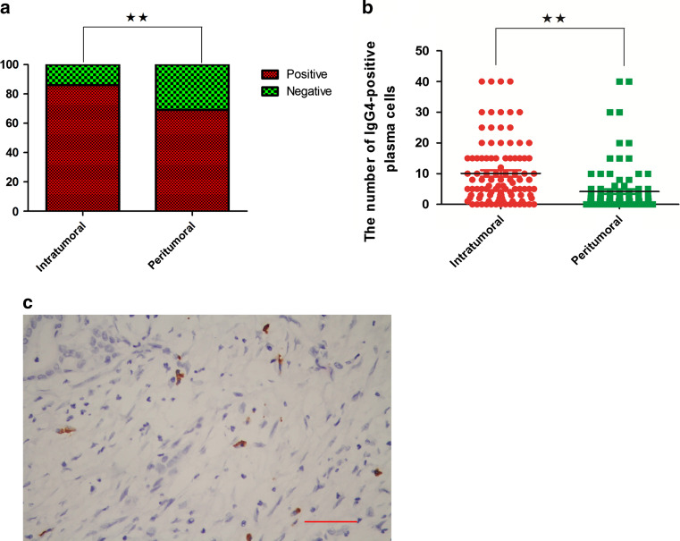
Fig. 1.
Distributions of IgG4-positive plasma cells in tumor and peritumoral tissues are different. a IgG4-positive plasma cells are found in 86 % of tumor tissue samples compared with 69 % of the peritumoral tissue samples (Chi-square test, **P < 0.01). b The infiltrations of intratumoral and peritumoral IgG4-positive plasma cell in each case are compared (Wilcoxon signed-rank test, **P < 0.01). c Representative figures of IgG4-positive plasma cells, which are immunochemically stained brown and had a large eccentric nucleus, are shown (the scale bar represented 200 μm, 200×, magnification)

