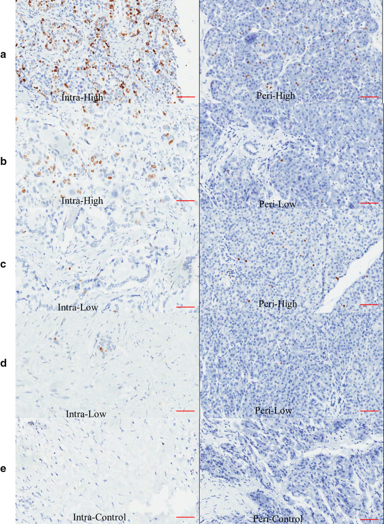
Fig. 3.
Representative figures of the different combinations of immunohistochemical staining of peritumoral and intratumoral IgG4-positive plasma cells are shown: a high-level infiltration of both intratumoral and peritumoral IgG4-positive plasma cells; b high-level infiltration of intratumoral and low-level infiltration of peritumoral IgG4-positive plasma cells; c low-level infiltration of intratumoral and high-level infiltration of peritumoral IgG4-positive plasma cells; and d low-level infiltration of both intratumoral and peritumoral IgG4-positive plasma cells. e Control staining of the tumor and peritumoral tissues. IgG4-positive plasma cells are immunochemically stained brown and had a considerable nucleus-to-cytoplasm ratio, as well as an eccentric nucleus (the scale bar represented 150 μm, 200×, magnification)

