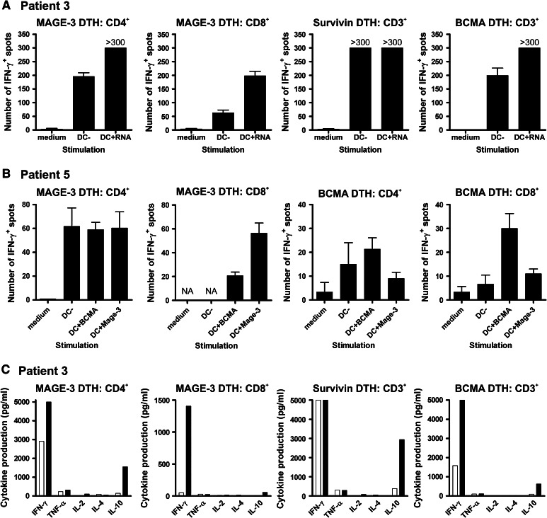
Fig. 3.
Cytokine release by TAA-specific T cells upon antigen recognition. DTH-infiltrating lymphocytes were cultured for 3-4 weeks in medium containing 200 U/mL IL-2 and 10 ng/mL IL-15. Thereafter, CD3+, CD4+ and/or CD8+ T cells were isolated and overnight restimulated with target cells. a–b IFN-γ production by TAA-specific T cells of patients 3 (a) and 5 (b) was analyzed using the ELISPOT assay. c Multiple cytokine levels were measured in culture supernatants of patient 3 using a cytokine bead array and flow cytometry

