Fig. 1.
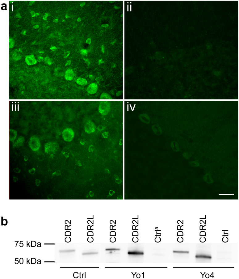
CDR2 and CDR2L antibodies in sera from Yo-positive patients. a shows sections of rat cerebellum with immunofluorescent staining of patient sera and rabbit CDR2 and CDR2L antibodies. Serum from Yo6 (i) stains the cytoplasm of Purkinje, stellate, and basket cells, similar to the rabbit CDR2L antibody staining (iii). Serum from CDR2L4 (ii) does not stain the sections, whereas the rabbit CDR2 antibody (iv) shows weak cytoplasmic Purkinje cell staining. Scale bar 50 μm. b western blot of recombinant CDR2 and CDR2L protein incubated with patient sera and rabbit CDR2 antibody and CDR2L antibody (Proteintech). Sera from Yo1 and Yo4 show bands coinciding with rabbit CDR2 and CDR2L antibodies of around 62 and 58 kDa, respectively, representing antibodies against CDR2 and CDR2L proteins in the patients sera. Control lanes with reticulocyte lysate without recombinant proteins are negative. aCtrl: control lane

