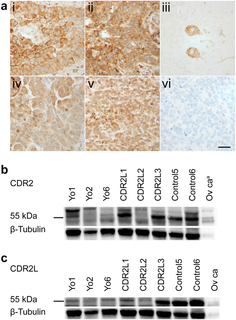
Fig. 2.
Expression of CDR2 and CDR2L proteins in human ovarian cancer. a shows formalin-fixed and paraffin-embedded sections of ovarian HGSC and cerebellum with immunostaining with rabbit CDR2 and CDR2L antibodies. Ovarian cancer sections from Yo4 (i) and CDR2L3 (ii) show strong CDR2 cytoplasmic staining of cancer cells and similar, but less intense, CDR2L staining (iv, v). In cerebellar sections, Purkinje cell cytoplasm shows CDR2L antibody staining (iii) and ovarian cancer section incubated with only secondary antibody shows no staining (vi). Scale bar 50 µm. b, c, western blots of human ovarian cancer lysates (50 μg protein) from patients Yo1-2, Yo6, CDR2L1-3, and Control5-6 were incubated with rabbit CDR2 antibody and CDR2L antibody (Sigma-Aldrich), respectively. The control lane to the right in both blots represents purchased ovarian cancer lysate (20 μg protein). In b, CDR2 protein presents as a double band around 55 kDa and an additional band at around 60 kDa in all samples. In c CDR2L presents as one band around 55 kDa in all lanes. Western blots were normalized to β-tubulin. aPurchased ovarian cancer lysate

