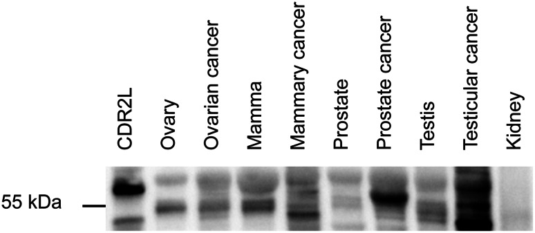
Fig. 3.
Expression of CDR2L protein in various human tissues. Western blot of lysates from various human tissues (20 μg protein) incubated with rabbit CDR2L antibody (Sigma-Aldrich). CDR2L protein presents as a single or double band around 55 kDa and an additional band around 60 kDa in all samples (very faint in normal kidney lysate). Recombinant CDR2L in the lane to the left serves as positive control and shows a band around 58 kDa

