
Fig. 2.
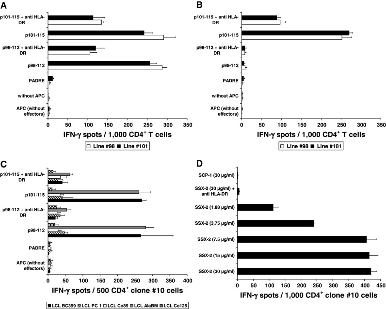
Dissection of the HLA-DR restriction of the T-cell response against SSX2-derived peptides p98-112 and p101-115 and natural processing and presentation of epitope p98-112. a Restriction of the T-cell response against p98-112 and p101-115 using the melanoma cell line SK-MEL-37 as APC. This cell line shares only DRB1*0101 with the BC399-derived T cells from line #98 and #101. DR restriction was demonstrated by blocking the response of both T-cell lines against APC pre-incubated with the anti-DR antibody clone L243. b DRB1*0701 restriction of the response from T cells of the BC399-lines #98 and #101 against LCL Co89 pulsed with peptide p101-115. *0701 is the only DRB1 subtype shared by Co89 and patient BC399. c Dissection of the restriction of the T-cell response of clone #10 from patient BC418: a response after stimulation with p98-112 or p101-115 only was observed when these peptides were presented by LCL from BC399 and AlaBM (P < 0.01) proving the restriction of these T cells to HLA-DRB1*0101, which is the only DR molecule shared by effector cells and APC. There was no response if both peptides were presented on LCL from PC 1, Co89, or Co125 excluding a restriction to HLA-DRB1*0301 or DRB3*0202. Y-axes of the A, B, and C panels display the different APC settings used to challenge T cells during IFN-γ ELISPOT. d Loading of autologous DC (1 × 10⁴/well) with increasing concentrations of SSX2 whole protein induced a concentration-dependent response of clone #10 CD4+ T cells, which was blocked by anti-HLA-DR. The control antigen (SCP1 fragment p630-817) was not recognized (P < 0.001). Y axis shows the different DC settings used to challenge T cells during IFN-γ ELISPOT