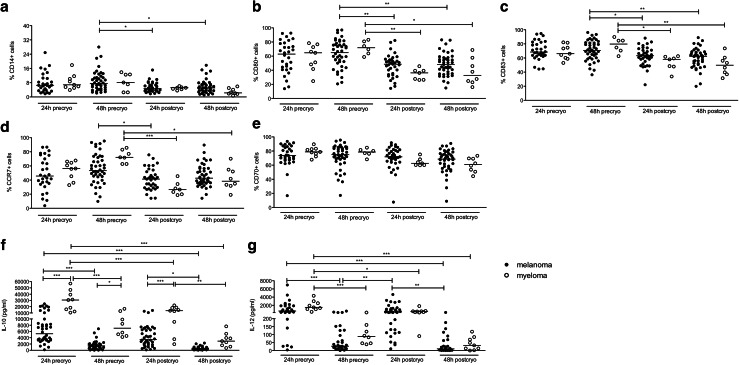
Fig. 4.
Quality of in vitro generated DCs from MM patients. Monocytes were isolated from PBMCs through plastic adherence and DCs were generated. On day 6, the immature DCs were harvested and electroporated with mRNA encoding TriMix1 and MAGE-C1. Twenty-four and 48 h after the electroporation, the DC phenotype was analyzed and supernatants were collected to measure cytokine secretion. In addition, part of the DCs was stored in nitrogen for quality control after a freeze/thaw cycle. The graphs show the phenotype of the DCs as determined by flow cytometry (panels a–e) and the DC cytokine production as determined by ELISA (panels f, g) from melanoma compared to MM patients 24 and 48 h after electroporation, either before (‘precryo’) or after (‘postcryo’) cryopreservation. Each symbol represents one experiment, and the median of all experiments is indicated by the line. All statistically significant differences are indicated: ***p < 0.001; **p < 0.01; *p < 0.05

