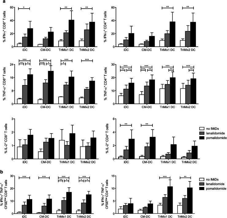
Fig. 5.
Costimulatory effect of IMiDs is enhanced when combined with TriMix DCs. T cells from MM patients were labeled with CellTrace Violet, stimulated with anti-CD3-coated microbeads. To some of the conditions, autologous iDCs, CM-DCs, TriMix1 DCs or TriMix2 DCs were added. After 6 days of T-cell stimulation, proliferation was measured and an intracellular flow cytometry staining was performed to evaluate the IFN-γ, TNF-α and IL-2 production by the CD4+ and CD8+ T-cell compartment. a The bar graphs indicate the mean percentage of cytokine-producing T cells, and the error bars indicate the SEM (n = 4). b The percentages of IFN-γ+ TNF-α+ cells within the CellTrace Violetlow (proliferating) CD8+ or CD4+ T-cell population are shown on the overview graphs. The bar graphs indicate the mean percentage of cytokine-producing T cells, and the error bars indicate the SEM (n = 4). TriMix1 = caTLR4 + CD40L + CD70, TriMix2 = caTLR4 + CD40L + 4-1BBL. Statistically significant differences resulting from the addition of IMiDs are indicated: ***p < 0.001; **p < 0.01; *p < 0.05

