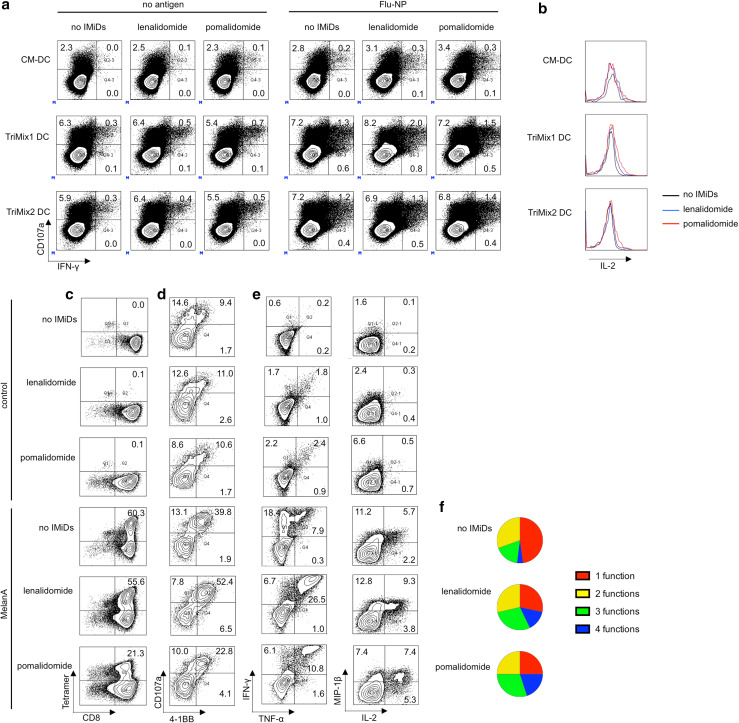
Fig. 6.
IMiDs enhance the quality of antigen-specific T cells stimulated or induced by TriMix DCs. a, b Purified CD8+ T cells from MM patients were stimulated with DCs electroporated with Flu-NP-encoding mRNA or electroporated without antigen-encoding mRNA (negative control). After 3 days, intracellular flow cytometry stainings were performed to evaluate the CD107a, IFN-γ and IL-2 expression by the CD8+ T cells. a Flow cytometry plots showing the percentages of IFN-γ+, CD107a+ and IFN-γ+ CD107a+ CD8+ T cells. b Expression of IL-2 in CD107a+ IFN-γ+ CD8+ T cells. Results in panels a, b are representative of two experiments. c–f Purified CD8+ T cells from healthy donors were stimulated with TriMix1 DCs co-electroporated with Melan-A encoding mRNA. After three rounds of stimulation, T cells were co-cultured with Melan-A or Gag (control) peptide pulsed T2 cells and the functionality of the Melan-A-specific T cells was examined. c Flow cytometry plots showing the percentage of Melan-A-specific CD8+ T cells. d Flow cytometry showing the percentage of CD107a+ 4-1BB+ Melan-A-specific CD8+ T cells. e Flow cytometry pots showing the percentage of cytokine-producing Melan-A-specific T cells. f Pie charts indicating the proportion of polyfunctional Melan-A-specific CD8+ T cells. Results in panel c–f are representative of at least two experiments

