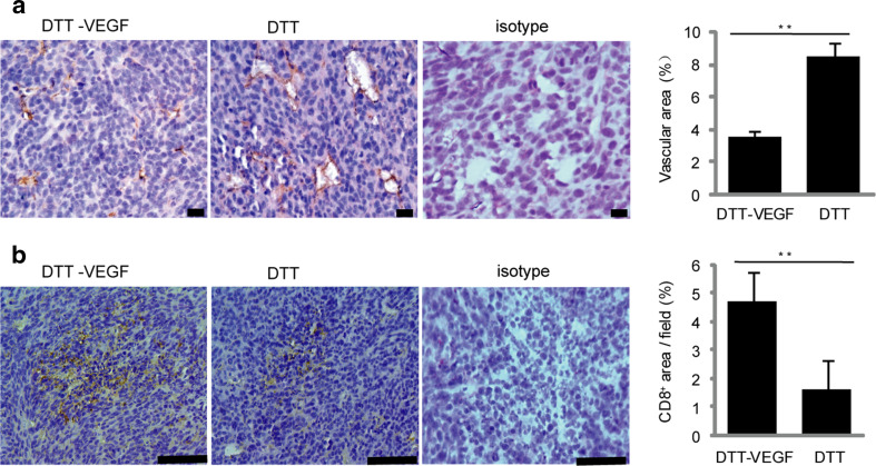
Fig. 6.
DTT-VEGF vaccine reduces tumor vascular growth and enhances T cell infiltration. CT26 tumors (n = 5) were collected at day 14 after tumor challenge as in Fig. 5d, sectioned and immunohistochemically stained. a Immunohistochemical staining of CD31+ cells. Scale bar 20 μm. Quantification of CD31+ blood vessels (mean % of CD31-covered area/field ± SEM) is shown in the bar graph on the right. b Immunohistochemical staining of CD8+ cells. Scale bar 100 μm. Quantification of CD8+ cells (mean % of CD8+-covered area/field ± SEM) is represented in the bar graph on the right. **p < 0.01, Mann–Whitney U test

