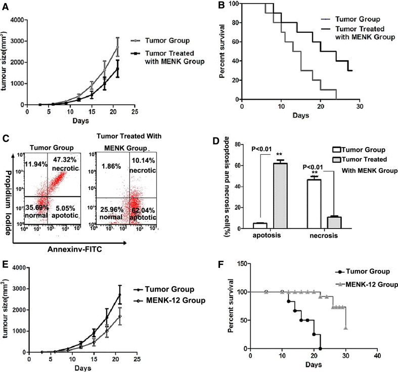
Fig. 5.
Antitumor effect by MENK in vivo. a–d The tumor models were established in C57BL/6 mice and then one group was treated with MENK (20 mg/kg) every other day for successive 30 days. Tumor sizes of each mouse (5/group) were measured every 3 days, and survival condition in each group (5/group) was monitored daily. The effect of apoptosis on tumor tissue of tumor group and MENK-treated group were analyzed. a Tumor sizes in the MENK-treated group showed significance. b Significant increase in survival rate and prolonged survival times were observed in MENK-treated group. c, d The apoptosis analysis of tumor tissue stained with both annexin V and propidium iodide (PI) by FCM. *p < 0.05 versus tumor group, **p < 0.01 versus tumor group. e, f Inhibiting effect on tumor growth by infusing differentiated BM-derived DCs into mouse. With use of 10−12 mol/L MENK, we got differentiated BM-derived DCs. Subsequently, we injected these differentiated BM-derived DCs into tumors bearing mouse. Tumor sizes and survival rate were measured separately. e Significant increase in survival rate in MENK-12 group compared with tumor group. f Tumor sizes in MENK-12 group showed significance compared with those in the tumor group. *p < 0.05 versus tumor group, **p < 0.01 versus tumor group

