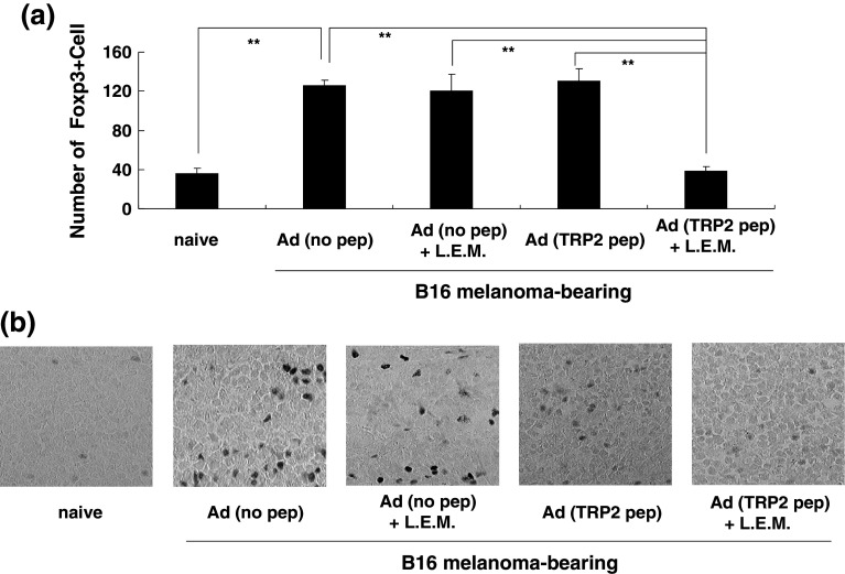
Fig. 5.
Immunohistochemical staining of Foxp3+ cells in draining LNs. a B16 melanoma-bearing mice were treated as described in Fig. 1. On day 21, draining LNs were harvested and stained with methyl green. Foxp3+ cells in five different fields were counted and the means ± SD are shown. **P < 0.01. b Representative photos. Foxp3+ cells are shown as black dots. Ad adjuvant

