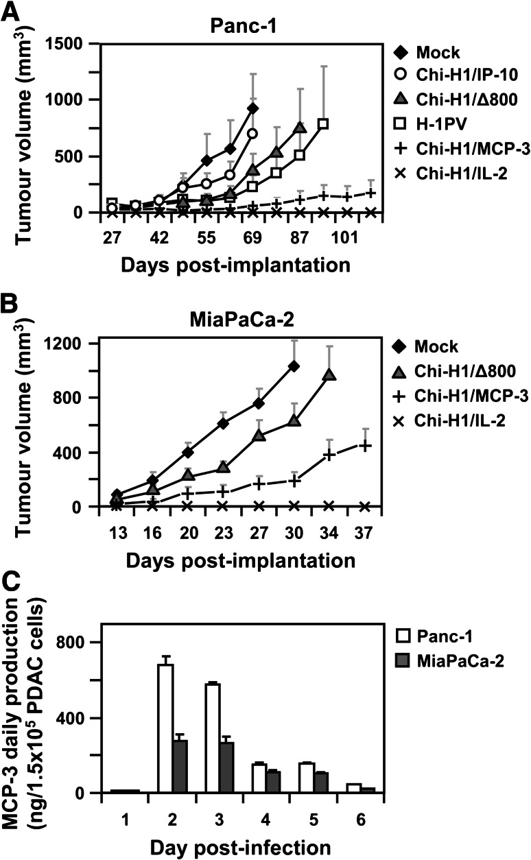
Fig. 4.
IL-2- and MCP-3-encoding H-1PV vectors suppress human PDAC tumour development. a 5 × 10⁶ Panc-1 cells were buffer-treated (Mock), infected at an MOI = 1.5 RU/c with wild-type H-1PV (H-1PV) or Chi-H1/-based vectors, empty (∆800) or transducing IP-10, MCP-3 or IL-2, and s.c. implanted into female Balb/c mice (n = 8, wild-type H-1PV: n = 5). The two-dimensional protrusions of the developing tumours were measured twice a week using an electronic calliper and tumour volumes calculated using the following formula: V = ½ × L × B2. Data represent mean tumour volumes ± SEM. b 2.5 × 10⁶ MiaPaCa-2 cells were buffer-treated (Mock) or infected (MOI = 2 RU/c) with Chi-H1/∆800, Chi-H1/MCP-3 or Chi-H1/IL-2, s.c. implanted into nude Balb/c mice (n = 10, Chi-H1/IL-2: n = 6), and tumours were monitored as described in a. c 1.5 × 10⁵ Panc-1 and MiaPaCa-2 cells were infected with Chi-H1/MCP-3 (MOI = 3 RU/c), and the daily secretion of MCP-3 was analysed in cell supernatants using specific ELISA. Data represent means ± SD of three measurements of a representative infection

