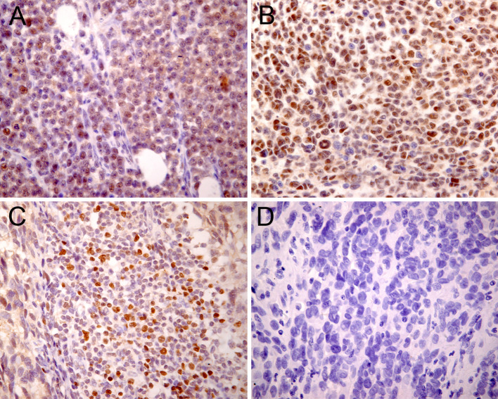
Fig. 1.
Immunohistochemical staining of paraffin-embedded melanoma tissues revealed Foxp3 expression in the melanoma cells. Nuclear/cytoplasmic (a) and nuclear (b) Foxp3 staining patterns were observed. Foxp3-positive lymphocytes (c) were probably tumor-infiltrating T regulatory cells (Tregs). In nonmalignant melanocytic nevus tissues, no Foxp3-positive cells were detected (d)

