
Fig. 4.

Effect of CD39, CD73 or A2A adenosine receptor inhibition on the suppressive capacity of OvCA cells against proliferating activated CD4+ T cells. a–f Treg and OvCA cells were co-cultured with CD4+ T cells that had been stained with CFDA-SE (a–e) or Cell Proliferation Dye eFluor® 670 (f) and stimulated with plate-bound anti-CD3 and anti-CD28 antibodies. To inhibit ectonucleotidases, CD39 or CD73 inhibitors ARL67156 (ARL) and APCP were added as indicated (100 μM each). ADORA2A was blocked using 100 nM SCH58261. To activate adenosine receptors, 10 μM adenosine-5′-N-ethylcarboxamide (NECA) was used. a During each cell division, CFDA-SE is distributed equally between the daughter cells, which enables the assessment of a cell’s proliferation history by flow cytometry. In order to determine the total number of cell divisions, the number of counts in each region was divided by 2n (n = number of peak, from right to left, beginning with 0) to determine the number of cells ‘x’ which originally divided into the cytometrically assessed cells of the peak. 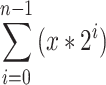 gives the total number of cell division per peak n. b–f Representative and independent experiments are shown (n = 3) for the co-culture of CD4+ T cells with Treg (b), SK-OV-3 and OAW42 cells only (c), with siRNA-transfected SK-OV-3 and OAW42 cells (d), with SK-OV-3 and OAW42 cells in the presence or absence of the inhibitors ARL67156, APCP or SCH58261, which block CD39, CD73 or ADORA2A, respectively, (e) and with SK-OV-3 and OAW42 cells in the presence of NECA ± ARL67156 or APCP or both (f)