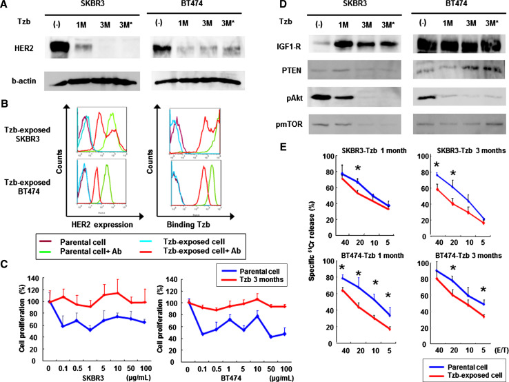
Fig. 1.
Effects of continuous exposure to trastuzumab in HER2-overexpressing breast cancer cells. a Western blot analysis of HER2 expression. Human breast cancer SKBR3 and BT474 cells were initially incubated with 50 mg/ml trastuzumab (Tzb) for 1 month followed by 100 mg/ml trastuzumab for 2 months. *Cells were cultured in the absence of trastuzumab for 5 days before analysis. Equivalent amounts of protein from whole cell lysates were loaded into each lane. Blots were probed with anti-HER2-ECD antibody and visualized by using an ECL detection system. Equal loading of samples was confirmed by stripping each blot and reprobing with anti-β-actin antibody. b Flow cytometric analysis of HER2 expression and trastuzumab binding. Parental or trastuzumab-exposed cells were stained with APC-conjugated anti-HER2-ECD antibody to measure cell-surface HER2 expression or treated with trastuzumab followed by incubation with APC-conjugated anti-human antibody to measure the amount of bound trastuzumab. c Parental or trastuzumab-exposed cells were further treated with the indicated doses of trastuzumab for 5 days, and cell viability was assessed by XTT assay. d Western blot analysis for assessment of HER2-related signaling pathway. Blots were probed with anti-IGF1-R, anti-PTEN, anti-phosphorylated Akt, or anti-phosphorylated mTOR antibody. e ADCC activity of trastuzumab-exposed SKBR3 or BT474 cells. Parental or trastuzumab-exposed cells were incubated with PBMCs from healthy donors in the presence of 10 μg/ml of trastuzumab, and the cytotoxic activity was assessed by a 4-h standard 51Cr-release assay. Data represent the mean ± SD of 3 wells at four different effector-to-target (E/T) ratios. *p < 0.05

