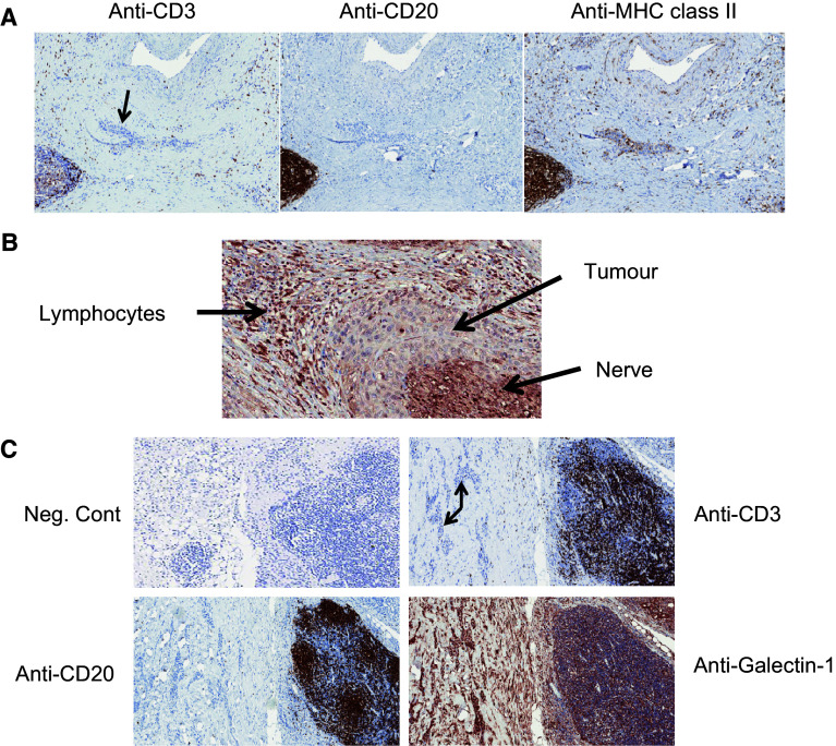
Fig. 2.
Immunostaining patterns for CD3, CD20, MHC II and galectin-1 in perineural SCC. a Consecutive tissue sections from a single patient were stained with antibodies directed against CD3 (left panel), CD20 (middle panel) and MHC class II (right panel). Tumour tissue is indicated by the arrow. Magnification ×9 for all images. b Galectin-1 staining from a single patient showing the relationship between lymphocytes, tumour and nerve tissue. Magnification ×18. c Consecutive tissue sections from a single patient were stained with antibodies directed against CD3, CD20 and galectin-1. Tumour tissue is indicated by the arrow. Magnification ×7

