Fig. 2.
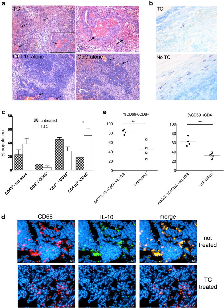
Leukocyte recruitment and massive necrosis characterize regressing tumors soon after combinatory treatment. a Histological analysis of tumor site at early time point after first treatment (48 h); hematoxylin/eosin staining for triple-combination treatment and single-agent treatment; black arrows indicate areas of necrosis; upper right panel shows an enlargement of image on the left (magnification ×10 and ×40 for the inset). b Representative toluidine staining of tumor samples at 48 h after first triple-combination treatment (upper panel) and untreated tumors (lower panel) showing mast cell infiltration (magnification ×40). c FACS analysis of leukocyte population in tumor samples at 48 h after first treatment of triple-combination therapy in comparison with untreated tumors. Mean of four tumors per group is shown; the experiment repeated twice. Antibodies for CD45, CD4, CD8 and CD11b have been used, and gating on CD45+ cells has been done to evaluate the percentage of the different leukocyte populations. d Representative double immunofluorescence staining of tumors 48 h after first triple-combination treatment (lower panels) and untreated tumors (upper panels) showing colocalization of CD68+ cells with IL-10 expression. Single-channel and merged images are shown. e FACS analysis of the activation state of CD8 and CD4 T cells in tumor samples at 48 h after first treatment of triple-combination therapy in comparison with untreated tumors. Percentage of CD69+ cells is done gating cells on CD8 and CD4 cells. For each analysis, at least four mice per group have been analyzed, and the experiment repeated three times

