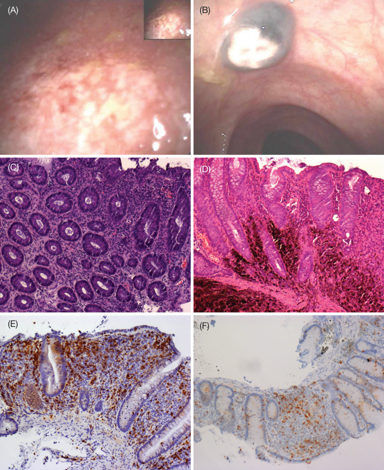
Fig. 5.
Colonoscopic images of a grade 3 ulcerative colitis with b evidence of colic uveal melanoma metastasis. Hematoxylin and eosin staining of biopsies demonstrates c autoimmune colitis with inflammatory cells, erosion of the tonaca propria and loss of gland goblet cells, and d metastatic deposits. Immunohistochemical labeling indicates e most of the inflammatory cells are of the CD8+ phenotype and f there is a decrease in CD8+ T-cells following steroid therapy. All images were captured at ×100 magnification

