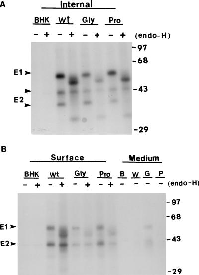
FIG. 4.
Cell surface expression of wild-type and mutant proteins. Induced BHK cells were labelled with [35S]methionine for 30 min and chased with 1 mM unlabelled methionine for 2 or 8 h. Cell surface RV antigens were derivatized with sulfosuccinimidobiotin and isolated as described in Materials and Methods. The virus spike proteins were precipitated and separated into internal (A) and surface (B) proteins by streptavidin binding. Portions of the immunoprecipitates were (+) or were not (−) digested with endo-H. The positions of apparent molecular mass markers are shown at the right (in kilodaltons). Internal, intracellular antigens; surface, cell surface antigens; medium, RV antigens released into the culture medium; Wt, W, BHK-E2E1; Gly, G, BHK-E2E1(G93D); Pro, P, BHK-E2E1(P104G).

