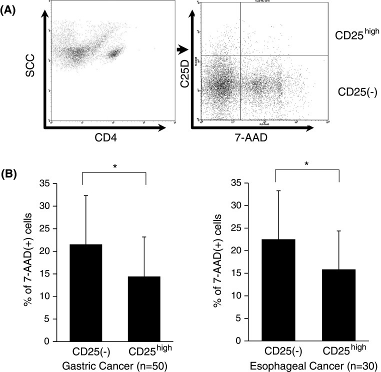
Fig. 3.
Reduced apoptotic cells in CD4(+)CD25high regulatory T cells. The proportion of apoptotic cells showing 7-AAD-positive staining between CD4(+)CD25(−)T cells and CD4(+)CD25highTregs was evaluated by flow cytometry (a). Summarized data derived from freshly isolated TILs in gastric and esophageal cancer are shown in b. Data were analyzed using a non-paired Student’s t-test, and findings were considered significant when p values were <0.01 (*)

