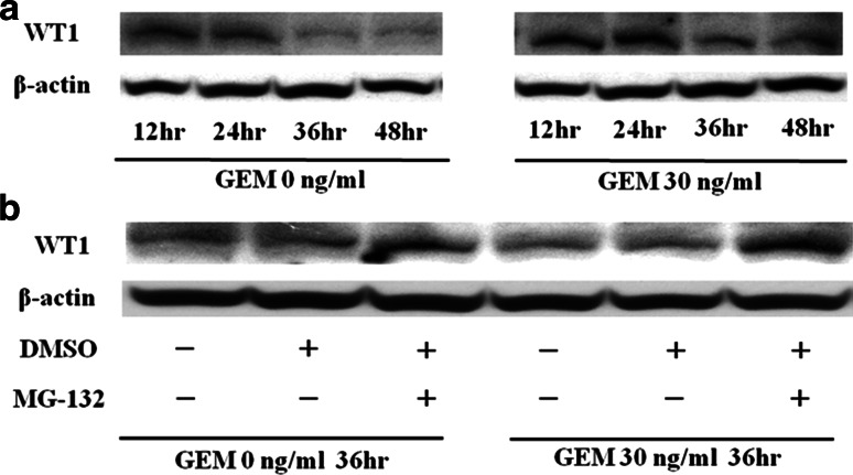
Fig. 4.
a WT1 protein is degraded by proteasomal enzymes. Twenty-four hours after 3 × 10⁵ MIAPaCa2 cells/well were seeded in 6-well culture plates, medium was exchanged from untreated to media containing GEM (0 or 30 ng/ml). Expression of WT1 protein in the cells was analyzed every 12 h thereafter from immunoblots described in Sect. “Materials and methods”. b Protease inhibitors block WT1 degradation. Twenty-four hours after incubating MIAPaCa2 cells with GEM (0 or 30 ng/ml), MG-132 in DMSO or DMSO alone was added to each well at a concentration of 5 μM and 0.05%, respectively. Treated and control cells (in 0.05% DMSO alone) were incubated for 12 h before harvesting cells for immunoblot analysis of WT1 and beta-actin proteins

