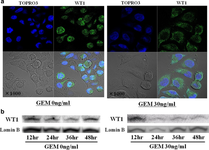
Fig. 5.
a GEM treatment shifts WT1 protein localization from nucleus to cytoplasm. Twenty-four hours after seeding 3 × 10⁵ MIAPaCa2 cells/well in 6-well culture plates, untreated medium was exchanged for fresh medium with or without GEM (0 or 30 ng/ml). After 24-h incubation, cells were fixed with paraformaldehyde, followed by nuclear staining with TO-PRO-3 iodide (blue color) and detection of WT1 with rabbit anti-WT1 polyclonal antibody and anti-rabbit IgG conjugated with fluorescein isothiocyanate (green color). Stained cells were observed using confocal microscopy (original magnification ×1,000). b GEM treatment diminishes nuclear localization of WT1 protein. Twenty-four hours after seeding 3 × 10⁵ MIAPaCa2 cells/well in 6-well culture plates, medium was exchanged for fresh medium with or without GEM (0 or 30 ng/ml). At 12-hour intervals thereafter, nuclei were isolated and WT1 protein levels of nuclear extracts were analyzed on immunoblots as described in Sect. “Materials and methods”

