Fig. 1.
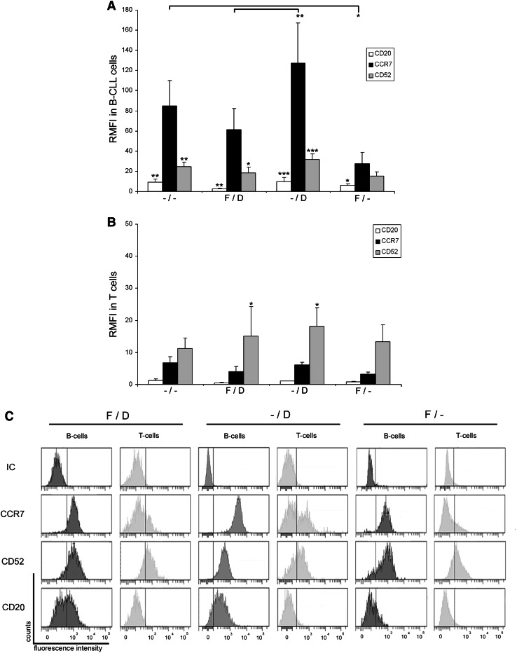
CCR7 surface expression in 17p-deleted and/or fludarabine-refractory CLL patients. a Relative median fluorescence intensity (RMFI) in CLL cells. CLL cells from peripheral blood samples (n = 20) were analyzed by FCM to determine CCR7, CD52 and CD20 surface density measured as median relative to a corresponding isotype control. Patients were distributed in four groups depending on their cytogenetic profile and fludarabine-refractory (FR) status: –/–, patients with normal cytogenetics and no previous treatment (n = 5); F/D, patients with 17p- and FR-CLL (n = 5); –/D, patients with 17p- and fludarabine-naïve CLL (n = 6); F/–, patients without 17p- and FR-CLL (n = 4). Bars represent mean ± standard mean error. b RMFI in T cells from CLL patient samples. CCR7, CD52 and CD20 were analyzed in T cells from the same blood samples as shown in a. c Frequency histograms showing CCR7, CD52 and CD20 in a representative patient of each group. The pattern and intensity of each surface marker is shown in both B and T cells. All statistical analyses in each group are referred to CCR7. *p < 0.05; **p < 0.01; ***p < 0.001

