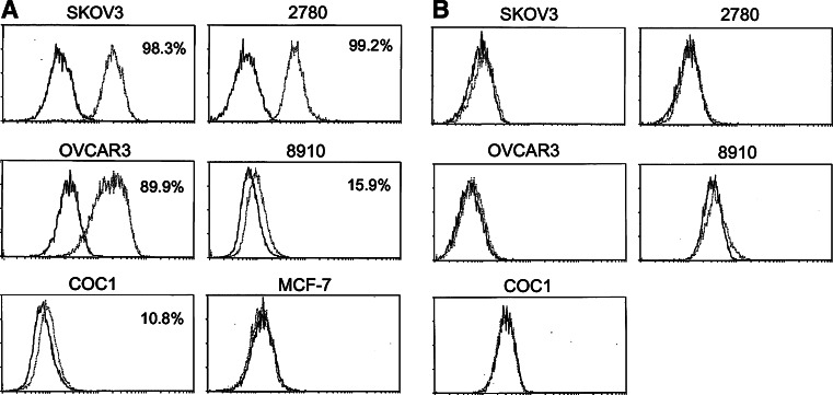
Fig. 1.
Expression of CD40 and CD40L on ovarian carcinoma cell lines. Cell surface expression of CD40 and CD40L on human ovarian carcinoma cell lines and breast carcinoma cell line (MCF-7) were studied separately using anti-CD40 (a) or anti-CD40L monoclonal antibodies (b) (open curve). Background staining is indicated (filled curve). The proportions of cells expressing the cell surface marker are indicated in each histogram

