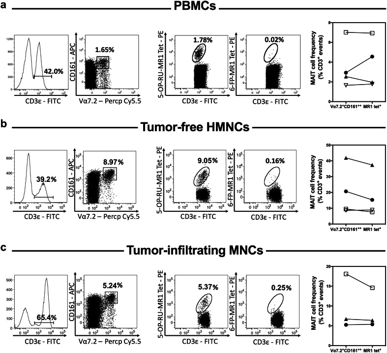
Fig. 1.
Peripheral blood and hepatic MAIT cells can be readily detected in CRC patients with liver metastasis. The frequency of MAIT cells was determined by flow cytometry among PBMCs (a), tumor-free, non-parenchymal hepatic mononuclear cells (b) and hepatic tumor-infiltrating mononuclear cells (c) isolated from patients with CRLM. MAIT cells were defined as CD3ε+Vα7.2+CD161++ or CD3ε+5-OP-RU-MR1 tetramer+ cells as indicated. 6-FP-MR1 tetramer was used as a staining control. Representative dot plots for each sample type are depicted (left panels). MAIT cell frequencies were calculated using both staining strategies for 3–4 patients (right panels). Values for matched samples (scatter plots with each patient being represented by a distinct symbol) are shown for comparison

