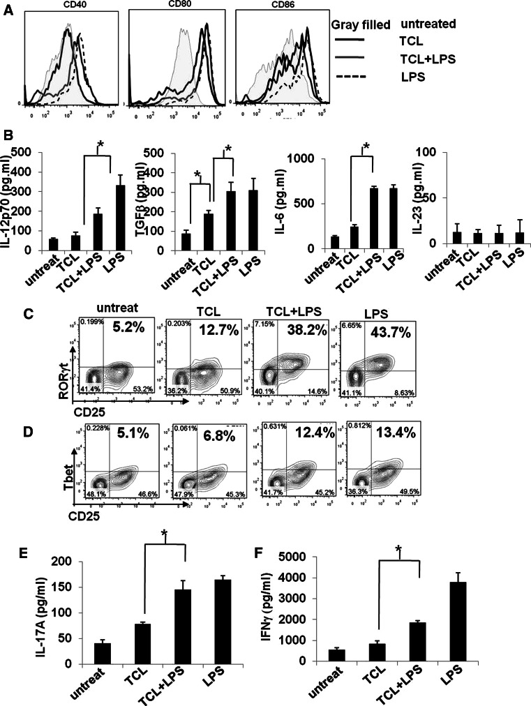
Fig. 3.
Treatment with TCL in combination with LPS activates DCs to full maturation and enhances the development of TCL-loaded DC-activated Th1/Th17. a Expression of cell surface markers (CD40, CD80, and CD86) on untreated DCs (gray-filled histogram), TCL-loaded DCs (black line open histogram), LPS-stimulated DCs (black-dashed line open histogram), and TCL-loaded plus LPS-stimulated DCs (gray line open histogram) were analyzed by flow cytometry and are presented as overlay histogram plot. An isotype-matched control mAb was used as control in all experiments (data not shown). b Expression of IL-12p70, TGFβ, and IL-6 in conditioned media from the co-culture of DCs and T cells was analyzed by ELISA. The effect of indicated DCs on the development of Th1/Th17 cells was evaluated by determining the levels of transcription factors RORγt (c) and Tbet (d) and of intracellular cytokines IL-17 (e) and IFNγ (f). * P < 0.05, between two indicated test groups. The results are representative of three independent experiments

