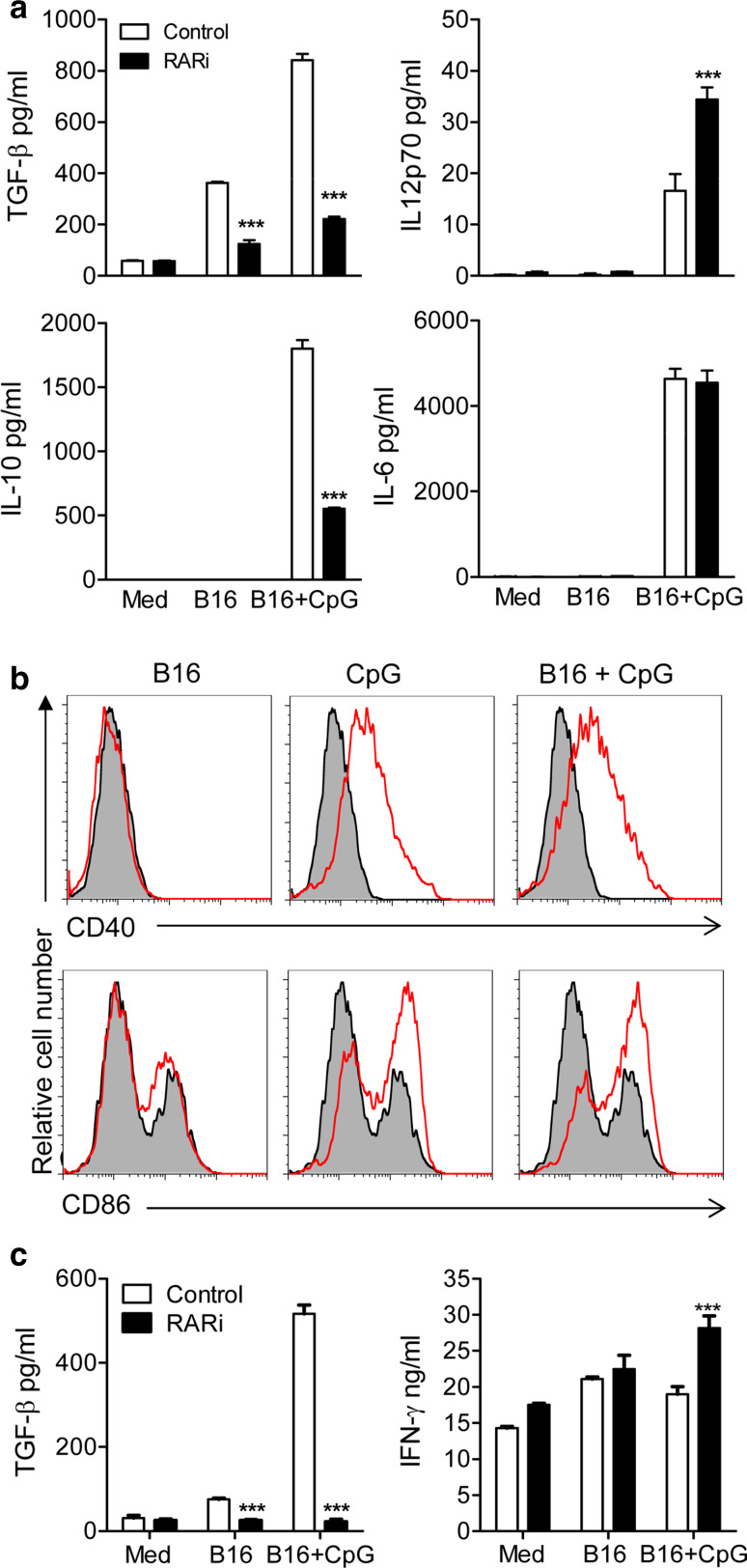
Fig. 4.
Inhibition of RA suppresses TGF-β and IL-10 production by DCs and induction of TGF-β-secreting T cells. a DCs were stimulated with the indicated combinations of hs/irr B16, CpG (5 μg/ml) and RARi. After 24 h in culture, TGF-β, IL-10, IL-12p70 and IL-6 concentrations were quantified in supernatants by ELISA. Results are means (±SD) of triplicate assays. Data are representative of six independent experiments. b DCs were stimulated with hs/irr B16, CpG (5 μg/ml) or both. After 24 h in culture, CD40 and CD86 expression was assessed by flow cytometry analysis. Results are presented as stimulated (red line) versus medium control (grey). c DCs were stimulated with the indicated combinations of hs/irr B16, CpG and RARi as in (a). After 24 h, DCs were washed and added to purified CD4+ T cells activated with αCD3 at a T cell: DC ratio of 5:1. After 3 days, supernatants were recovered and concentrations of TGF-β and IFN-γ were determined in triplicate assays by ELISA. Results are mean ± SE values for triplate assays and are representative of three independent experiments. ***p < 0.001 medium control versus RARi by Student’s t test

