Fig. 6.
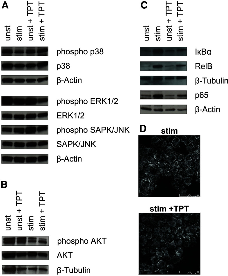
Topotecan affects intracellular signaling pathways in DCs. Protein samples of DCs generated as described were separated on SDS–PAGE, transferred to nitrocellulose membrane, and analyzed by immuno-blotting for the content of signaling proteins. Total and phosphorylated a p38, ERK1/2, SAPK/JNK, b AKT, and c IκBα, RelB, and NF-κB were detected with specific antibodies each. a–c Graphs are representative for two to three independent experiments each. d For confocal microscopy cells (105) were spun onto glass slides. RelB was detected with specific antibodies, and intracellular distribution was analyzed using a CLSM as described in the methods section. Each picture is representative of five taken per sample. Pictures are representative for two independent experiments

