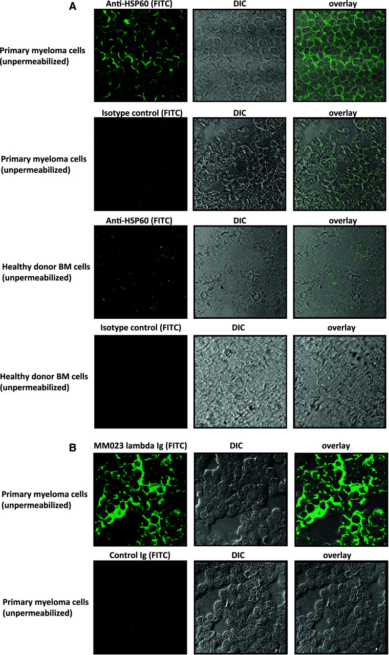
Fig. 5.
Surface exposition of HSP60 in primary myeloma cells. a HSP60 is displayed on the surface of non-permeabilized primary myeloma cells as visualized by confocal microscopy. Primary myeloma cells from bone marrow as well as bone marrow cells from healthy controls were spun on a slide and fixed with ethanol without further permeabilization. In the middle, the myeloma cells are shown in differential interference contrast (DIC). A HSP60 antibody or a murine isotype control antibody were used to stain the outer surface of the cells followed by secondary detection with an anti-mouse FITC-conjugated antibody (green) as shown in the left panels. The right panels display the overlay of both images. Images were obtained by confocal microscopy (Leika TCS SP2 AOBS; lens × 63) and analyzed using Leika confocal software. b Patient-derived HSP60-specific serum immunoglobulins recognize surface HSP60 on primary myeloma cells. Primary myeloma cells from bone marrow were spun on slides and fixed as described in a. The panels show (from left to right) primary myeloma cells stained with the lambda immunoglobulin fraction from patient MM023 or control IgG immunoglobulin followed by secondary detection with an anti-human FITC-conjugated antibody (green), myeloma cells in differential interference contrast and the overlay of both images. Images were obtained by confocal microscopy (Leika TCS SP2 AOBS; lens × 63) and analyzed using Leika confocal software

