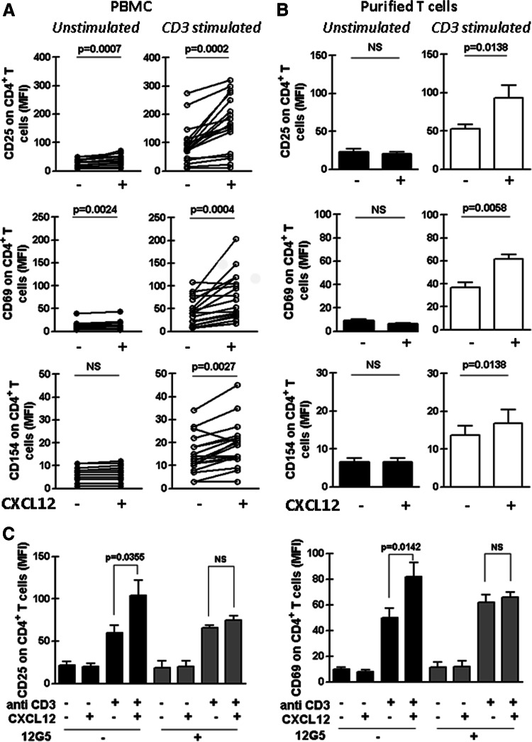
Fig. 1.
CXCL12 increases the expression of CD25, CD69 and CD154 on CD3-stimulated CD4+ T cells from CLL patients. PBMC from 18 CLL patients (a) or purified T cells from 9 CLL patients (b) were pre-treated or not with CXCL12 (1 μg/ml) for 2 h and then transferred to a well with immobilized anti-CD3 mAb (CD3 stimulated) or the corresponding isotype control antibody (Unstimulated). After 24 h of culture, CD25, CD69 and CD154 expression on CD4+ T cells was analyzed by flow cytometry. The figure shows the values of the mean fluorescence intensity (MFI) of CD25, CD69 and CD154 on CD4+ T cells of each patient obtained in PBMC cultures (a) or with purified T cell (b). c PBMC from 8 CLL patients were pre-treated anti CXCR4 mAb (12G5) or the corresponding isotype control antibody for 1 h before the co-stimulation assay. The bars represent the mean values ± SEM for the MFI of the activation marker on CD4+ T cells. The p values for the statistical analysis with the Wilcoxon’s signed rank test are shown in the figure. NS not statistically significant

