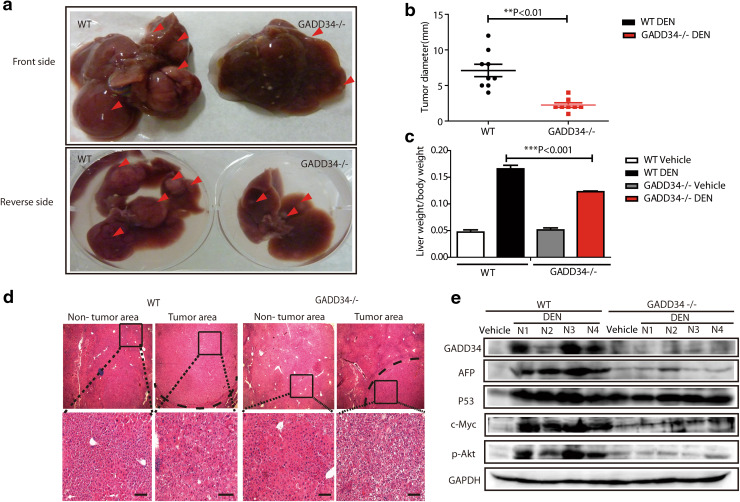
Fig. 4.
GADD34 deficiency reduced hepatocellular carcinoma (HCC) progression after DEN and TCPOBOP treatment. a Representative images with front and back side of HCC from WT and GADD34−/− mice. Red arrows indicate tumor nodules. b Statistical analysis of average tumor size. N = 9 for WT, n = 7 for GADD34−/−. **P < 0.01 versus WT. c Statistical analysis of liver/body weight ratios. ***P < 0.001 versus WT. d H&E staining of non-tumor and tumor liver sections obtained from WT and GADD34−/− mice. The outlined areas are enlarged in the lower panel. Scale bars 100 μm. e Analysis of indicated protein expression by Western blotting using whole-liver samples including tumor and non-tumor areas from WT and GADD34−/− mice

