Fig. 5.
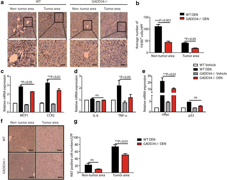
GADD34 deficiency reduced HCC progression through attenuating macrophage infiltration, cytokine level, oncogene expression and hepatic proliferation. a Representative IHC images of non-tumor and tumor areas in liver samples from WT and GADD34−/− mice using anti-F4/80 antibody. The outlined areas are enlarged in the lower panel. Scale bars 50 μm. b Statistical analysis of F4/80-positive cells from different locations in different samples. N = 6 with >8 fields of view for each group, *P < 0.05, ***P < 0.001 versus WT, mean ± SEM. c–e Real-time PCR analysis of indicated genes in non-tumor and tumor areas from WT and GADD34−/− mice. N = 4 for each experiment. *P < 0.05, **P < 0.01 versus WT, mean ± SEM. f Representative IHC results of Ki67 expression in both non-tumor and tumor area. Scale bars 100 μm. g Statistical analysis of Ki67-positive cells in (f). N = 6 with >8 fields of view for each group, **P < 0.01 versus WT, mean ± SEM. no significance versus WT

