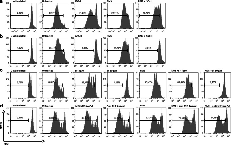
Fig. 5.
Proliferation of T cells during incubation with RMS culture medium and MIF inhibitors. a–d PBMCs were stimulated with IL-2 and OKT3 and cultured as indicated for 5 days. Proliferation was detected by a CFSE assay. Incubation with RMS supernatant (A204) slightly decreased the proliferation rate of T cells in each culture. No defects in the proliferation of T cells were observed during treatment with low concentrations of SF (3 µM), ISO-1, or anti-MIF antibodies, whereas higher concentrations of SF and Ant.III prevented T cell proliferation

