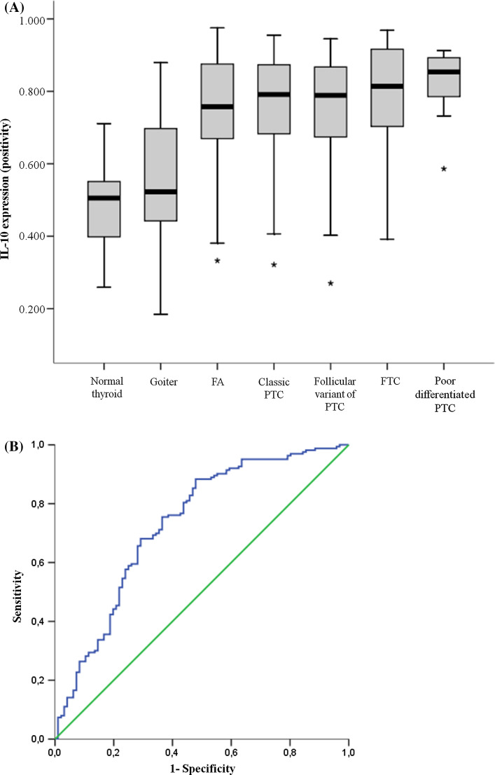
Fig. 4.
a Box-plot of IL-10 positivity of all histological sets of our samples. Note that normal thyroid presents the lowest IL-10 positivity, followed by goiters. These two tissues differ significant from all other pattern of thyroid lesions, including follicular adenomas and thyroid carcinomas. b ROC curve of IL-10 positivity, considering malignancy as positive. Abbreviations: FA follicular adenoma, FTC follicular thyroid carcinoma, PTC papillary thyroid carcinoma

