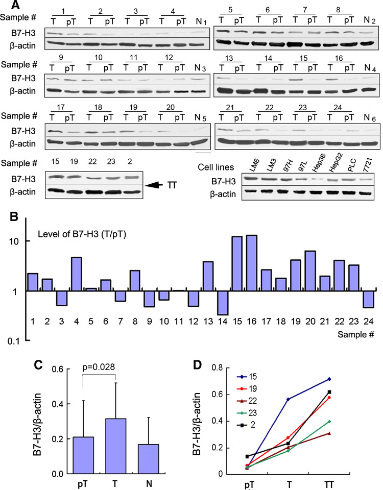
Fig. 2.
Western blot analysis of B7-H3 expression in HCC tissues and cell lines. a Western blot detection of 4Ig-B7-H3 protein in 24 paired tumors (T) and peritumoral tissues (pT) (including five cases with tumor thrombi [TT]), eight HCC cells lines, and six normal liver tissues (N). β-Actin served as a loading control. b The bar graph illustrates the ratio of the B7-H3 protein level (T/pT). Seventeen of 24 tumors (70.8 %) display upregulated B7-H3 protein levels, compared with the corresponding peritumoral liver tissues. c The B7-H3 protein level obtained by densitometric scanning is significantly upregulated in tumors compared with peritumoral tissues. d B7-H3 expression shows a sequential increase from peritumor tissues to tumor tissues, and to the corresponding tumor thrombi

