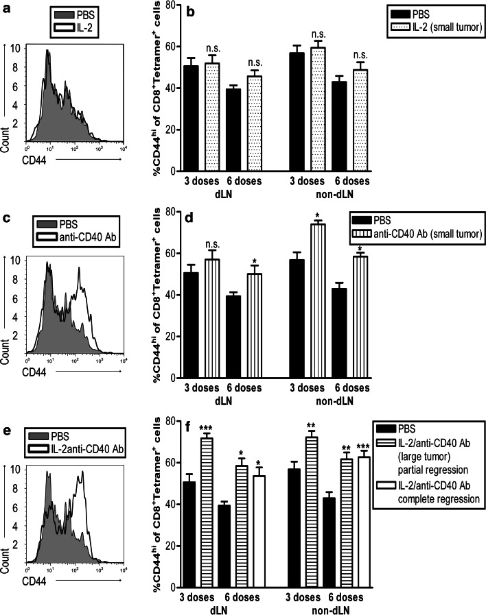
Fig. 3.
CD40 drives activated CD44hi tumor-specific CD8+ T cells. C57BL/6J mice bearing small (9–20 mm2) or large AE17-sOVA tumors (25–42 mm2) were given three or six i.t. doses of PBS (n = 8 mice), IL-2 (into small tumors, n = 8 mice; a, b), anti-CD40 Ab (into small tumors, n = 8 mice; c, d), or IL-2/anti-CD40 Ab (into large tumors, n = 10 mice; e, f). The dLNs and non-dLNs were prepared as single suspensions and stained for CD44, OVA-tetramer, and CD8. The percentage of OVA-tetramer+CD8+ cells that were CD44hi was calculated by flow cytometry. Representative histograms of CD44 staining from IL-2-treated (a), anti-CD40 Ab-treated (c) and IL-2/anti-CD40 Ab treated (e) from the dLN are shown after 6 doses. Pooled data (b, d, f) are shown from two experiments (42 mice for each timepoint, total mice = 84) represented as mean ± SEM. ***P < 0.001, **P < 0.01, *P < 0.05, n.s. not significant comparing treated groups to PBS-treated controls

