Fig. 1.
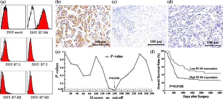
The monoclonal mouse anti-human B7-H4 antibody 3C8 was established and used in the immunohistochemical assay. a Flow cytometry analysis showed that the antibody specifically stained 293T-B7-H4 transgenic cells, and the staining was absent in the cross-reactions with 293T-mock cells and other B7 family molecules transgenic 293T cells. b Immunohistochemistry showed that positive B7-H4 immunochemical staining was predominantly observed on the membrane and in cytoplasm of tumor cells. c Negative B7-H4 immunochemical staining in normal esophageal tissue. d Negative control. e The minimum P value seek in the log-rank survival analysis of B7-H4 expression in ESCC tissues was performed, and when the cutoff value of H-score = 160 was selected, the minimal P value = 0.0105 was found. f The log-rank survival analysis was performed when H-score = 160. Scar bar = 100 μm

