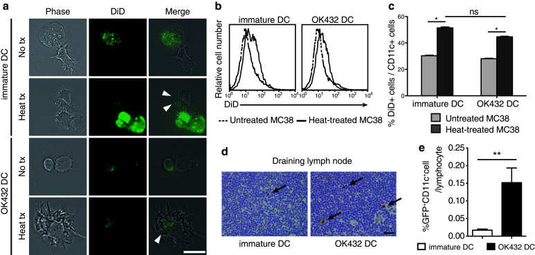
Fig. 1.

Effects of OK-432 on murine bone marrow-derived DCs. a OK-432-stimulated DCs or immature DCs were co-incubated for 3 h with MC38 cells untreated or treated at 80 °C for 90 s after staining with DiD dye. After incubation, DC and MC38 cells were observed using a fluorescence microscope. Arrowheads indicate MC38 derivatives being phagocytosed by DCs. No tx, untreated MC38 cells; heat tx, heat-treated MC38 cells; bar, 20 μm. b, c Co-incubated MC38 cells and DCs were stained with anti-CD11c antibodies and analyzed using flow cytometry. The histograms show the DiD fluorescent intensity of the CD11c-positive fractions. The percentages of DiD+ CD11c+ cells in the CD11c+ cell population are also shown in a column graph. The experiments were performed five times, and representative results are shown. Data are presented as the mean ± SE. *P < 0.05. d The migration abilities of the DCs after intratumoral transfer were evaluated. The draining lymph nodes were harvested at 3 days after RFA followed by the DC transfer. Frozen sections were prepared and stained with anti-GFP antibodies. Arrows indicate the GFP-positive cells in the lymph nodes. Bar 20 μm. e The draining lymph nodes were also analyzed using flow cytometry after staining with anti-CD11c antibodies. Data were obtained from six mice in each group. Percentages of GFP+ CD11c+ cell are presented as the mean ± SE. **P < 0.01
