Fig. 1.
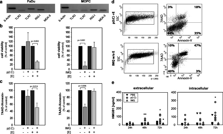
Poly(I:C) induces cell death in HNSCC via intracellular delivery and imiquimod via extracellular delivery. a Qualitative PCR analysis of unstimulated FaDu and MOPC cells shows basal expression of TLR 3, TLR 7, RIG-I and MDA-5; ß-Actin was used as a loading control. Shown is one representative example of two. b FaDu cells were stimulated with poly(I:C) [p(I:C)] or imiquimod [IMQ] for 72 h. Stimuli were delivered extracellularly (endocytic pathway) or intracellularly (cytosolic pathway, [E] = electroporation). Cell viability was analyzed by MTT assay. Unstimulated tumor cells were set as 100 %. The mean values ± SD from three independent experiments are shown. P value (t test) is indicated. c Cells were stimulated as in (b). Dead cells were identified by PE-conjugated annexin-V and 7-AAD staining and analyzed by flow cytometry. Unstimulated tumor cells were set as 100 %. The mean values ± SD from three to six independent experiments are shown. P value (t test) is indicated. One representative FACS-analysis of stimulated FaDu cells is shown in (d). e FaDu cells were stimulated with PBS (filled square), poly(I:C) (filled circle) or imiquimod (open triangle). Supernatants were collected after 24, 48 and 72 h, and HMGB1 concentration was analyzed by ELISA. Scatter plots depict seven independent experiments. P value (t test) is indicated

