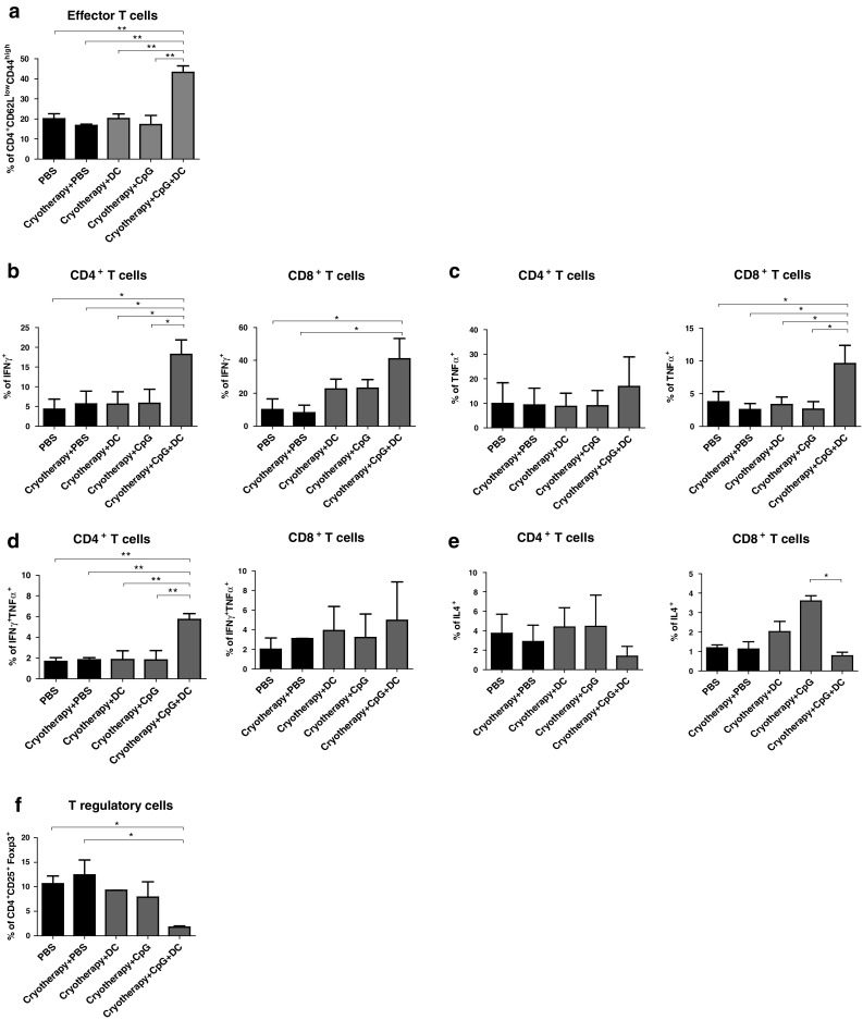
Fig. 4.

Immunocyte populations in the dLNs of tumor-bearing mice following treatments. Mice were inoculated i.f.p. with D122-luc-5.5 and further treated as described in materials and methods. Eight days post-second treatment, the dLNs were excised and analyzed by flow cytometry. a Percentages of CD4+ effector T cells (CD62Llow CD44high). b–e Percentages of CD4+ and CD8+ T cells secreting IFNγ (b), TNFα (c), IFNγ and TNFα (d), IL4 (e). f Percentages of Treg (CD4+CD25+Foxp3+) cells. Data presented as mean ± SEM, combined results of three independent experiments (n = 5–8 mice per group per experiment). *P < 0.05; **P < 0.001
