Fig. 3.

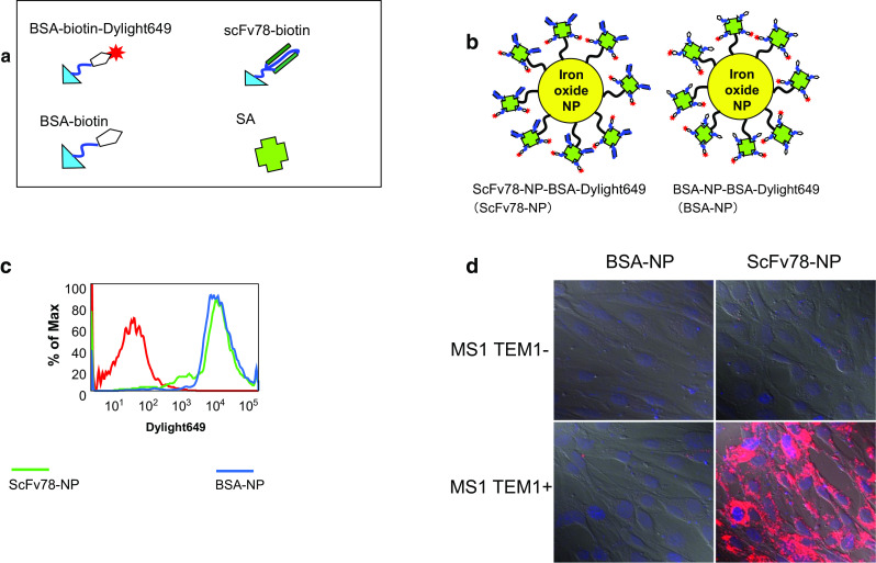
ScFv78-mediated NP internalization. a Illustration of the components used to prepare scFv78-NP-dye and BSA-NP-dye. b Streptavidin-conjugated iron-oxide nanoparticles co-labeled with BSA and BSA-Alexa649 (BSA-NP) or scFv78 and BSA-Alexa649 (scFv78-NP). c FACS analysis of the fluorescent intensity of the non-labeled nanoparticles (red), BSA-NP (blue) and scFv78-NP (green). d scFv78-NP or BSA-NP was incubated with MS1 and MS1-TEM1 (TEM1+) cells, respectively, in complete medium, and then internalization was analyzed by confocal imaging
