Fig. 2.
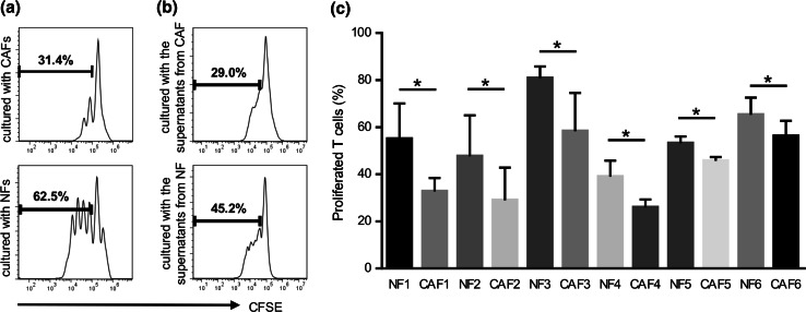
Suppressive activity on T cell proliferation by CAFs and NFs. Generated CAFs and NFs were co-cultured with CFSE-labeled T cells for 4 days with an anti-CD3/anti-CD28 stimulus. They were stained with APC-CD3 and 7-amino-actinomycin D (7-AAD) to gate viable CD3+ T cells. The proliferation of T cells was analyzed by the reduction of CFSE staining intensity using flow cytometry. a Representative data of the proliferation of T cells cultured with CAFs or NFs. b Representative data of the proliferation of T cells cultured with supernatants from CAFs or NFs. c The culture supernatant from CAFs showed greater suppressor activity than that from NFs in six HNSCC patients tested. Bars indicate mean values derived from 6 independent experiments. Asterisk indicates significant difference (P < 0.05) between NFs and CAFs

