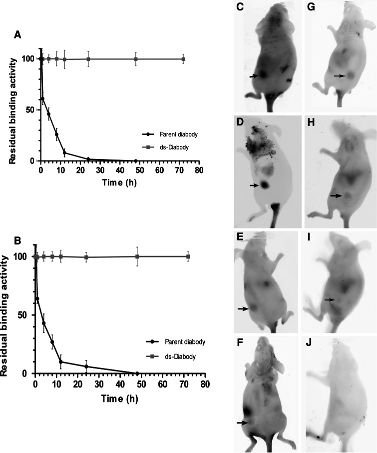
Fig. 3.
The in vitro and in vivo stability of the ds-Diabody in comparison with the parent diabody. Serum stability of the anti-Pgp-scFv component of the ds-Diabody and the parent diabody (a). Stability of the anti-CD3-scFv component of the ds-Diabody and the parent diabody (b). All data are normalized to time-point t 0 = 100%. Data shown are the mean percentage of residual binding ± SD of three independent experiments. Comparison of tumor localization between the ds-Diabody and parent diabody. Tumor localization by the ds-Diabody (c–f). Tumor localization by the parent diabody (g–j). The images were taken 2 (c, g), 12 (d, h), 24 (e, i), and 72 h (f, j) post-intravenous injection

